|
|

The CSPCA Charitable Trust needs your support through donations to continue vital research projects. Your donation is tax deductible.
RASP (Rescue A Shar-Pei) is a volunteer group with a concern for abandoned and abused Shar-Pei in Illinois / Indiana / Wisconsin and surrounding areas. |
 
|
 |
|

Health Issues
 Masticatory Myositis Masticatory Myositis
Masticatory mysositis appears to be a breed problem in the Chinese
Shar-Pei. While I'm hesitant to say it's an inherited condition I have
seen it in lines and in litters of affected individuals. This condition
used to be described as two separate disorders: (1)eosinophilic myositis
and (2) atrophic myositis. It is currently felt that these are
manifestations of the same disease now called masticatory myositis.
This is an inflammatory muscle disease, most likely immune-mediated,
involving the muscles of mastication - these muscles are used to grind
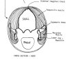
and chew food prior to swallowing. Five muscle groups in the dog are
involved in the process of mastication - four muscles are responsible
for closing the mouth and one with opening the mouth. The
tempoallis muscles and the masseter muscles are primarily the muscles
used to powerfully close the jaws and are espacially well developed in
Shar-Pei (Figs. 1 and 2).
Bear in mind that these muscles must also relax in order for the mouth to open.
Since these two muscles are the largest muscles which close the jaw, when they
become involved in masticatory myositis, the primary clinical sign is trouble in
opening the jaws.
Masticatory myositis can be divided into an acute and chronic
form. It must be remembered that one acute attack can lead immediately into
the chronic form of the disease, although, more often than not, multiple,
recurrent acute attacks are necessary. Symptoms of the acute form
involve the the swollen, firm and painful temporalis and masseter muscles.
The dog's head often appears swollen and larger than normal. The dog will
be reluctant or unable to open his mouth. Opening the mouth more than 1 inch
or so elicits an extremely painful response. This results in difficulty in
eating and often the owner notices increased drooling as well. This swelling
may even cause exophthalmia or the eyes to "bug out". Usually a fever is
present and the lymph nodes in the head and neck region are enlarged.
The tonsils are often enlarged as well, but it is difficult to visualize them
due to the inability to open the mouth. The patient is often depressed and
may resent palpation of the head musculature.
Laboratory findings are variable, but very often are normal. The white
blood cell count may be elevated and often there is an increase in eosinophils
(a type of white blood cell often seen with inflammation). Most often there
is a dramatic increase in a skeletal muscle enzyme known as creatine kinase
or creatine phosphokinase (CPK-MM). Smaller amounts of this enzyme
are also located in the brain (CPK-BB) and in the heart muscle (CPK-MB). This
enzyme has a short life span in the serum and is most often elevated in the acute form
of the disease due to the magnitude of muscle damage and because the owner usually
presents the dog while the disease is present. CPK may not be part of the normal
serum enzyme panel your veterinarian uses and may have to be requested separately.
Your vet should also request the CPK enzyme be reported in terms of the various
isoenzymes (heart, brain and skeletal muscles).
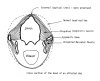
The chronic form usually is evident when the dog's head appears "sunken",
especially the top of the head. Sever and recurrent muscle damage leads to scar
tissue formation and atrophy or shrinking of the muscle. This scar tissue is non-functional
and these dogs often cannot open their mouths more than 1/2-1 inch. The dog's
head often appears "skull-like" with a prominent external sagittal crest
(the bony ridge on top of the head) and the eyes are sometimes enophthalmic or
sunken due to loss of the muscle mass behind them (fig.3). In the chronic
form, laboratory findings are often normal. The CPK-MM is usually normal due to the
fact that there is little muscle left to produce the enzyme. The dogs are normal
otherwise and non-painful, although the mouth cannot be opened.
The cause of masticatory myositis is uncertain, but is thought to be
immune-mediated because:
- The type of cellular infiltrate in the affected muscles.
- The disease is responsive to immunosuppressive doses of corticosteroids.
- In some cases, autoantibodies are present, fixed to the unique
muscles fibers present in the muscles of mastication (Type II M fibers)
and the presence of anti-Type II M antibodies in the serum of some
dogs with the disease. These antibodies may play a role in the immune
system attack on these muscle fibers.
Definitive diagnosis of this condition is based on muscle biopsy --
usually of the temporalis and/or masseter muscles. Also a 2M Antibody
test is available which can be done utilizing a serum sample -- this must
be done before therapy is initiated! This test available from:
Comparative Neuromuscular Laboratory
Basic Science Building, Room 1057
University of California, San Diego
La Jolla, CA 92093-0612
Phone: (858) 534-1537
Fax: (858) 534-7319
Treatment invariably involves the use of corticosteroids at high (immunosuppressive)
doses. Prednisolone is usually preferred. In the acute form, there is usually rapid
clinical improvement. The dose is subsequently reduced gradually and in some dogs,
prone to relapses, must be maintained on continuous alternate day therapy. In the
chronic form the prognosis is much more guarded. Surgery is usually done to allow
some return of jaw function. Often the insertion of the temporal muscle on the lower
jaw is surgically incised and released. This may free up the jaw enough for the dog
to be functional.
Fortunately, this is not a common disease, but one that veterinarians and owners
need to be aware of. |
|

))
))
))